
Field Notes · July 26, 2026 · 6 min · By Ximena Calloway
Reading Repigmentation: What the Pattern Inside Your Patch Predicts
Color coming back as tiny dots scattered across a patch and color creeping in from the edge are two different biological events with two different prognoses, and the difference is visible to you months before it is obvious to anyone else.
Treatment for pigment loss is slow in a way that is genuinely difficult to live with. Phototherapy is measured in months of appointments. Topicals are measured in months of application. And the standard follow up question, is it working, is usually answered with a shrug and a suggestion to keep going and see.
There is more information available than that, and it is sitting in your own patches. Repigmentation does not simply happen. It happens in patterns, the patterns correspond to which population of pigment producing cells is responding, and those populations have meaningfully different prognoses. A patch that is repigmenting in one pattern is telling you something different from a patch repigmenting in another, and you can see it long before the patch closes.
The original element in this piece is a monthly self photography protocol paired with a four pattern reading key, mapping what you see to which melanocyte reservoir is responding and therefore to what the next months are realistically likely to bring. Clinicians read these patterns routinely. Patients are almost never taught to, which is odd given that patients are the ones looking at the patch daily.
The biology in short. Repigmentation draws on more than one source. The best studied is the reservoir of melanocyte precursors sitting in the outer root sheath of hair follicles inside the patch. When stimulated, those cells migrate up and outward from each follicle, which produces the characteristic appearance of small pigmented dots that gradually enlarge and merge. A second source is the normally pigmented skin at the border of the patch, from which melanocytes advance inward, producing a patch that shrinks from its edges. Work combining topical calcineurin inhibitor therapy with narrowband ultraviolet B has demonstrated activation of both follicular and dermal melanocyte precursor populations histologically as well as clinically (Archives of Dermatological Research, 2021).
Those two mechanisms behave differently, and that is the whole practical point.
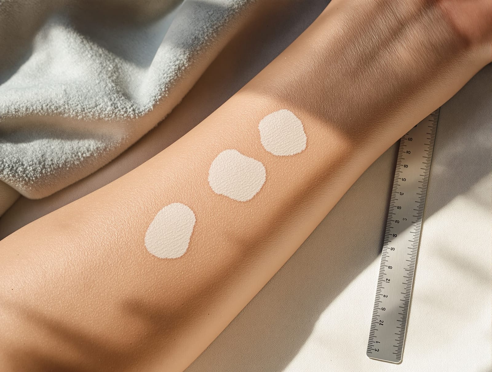
The photography protocol. Once a month, on a fixed date, photograph each patch you are tracking under these conditions. Daylight from a window, never a flash, and never overhead bathroom lighting, which flattens the exact contrast you are trying to see. The same room and roughly the same time of day each time. Camera at a consistent distance, ideally with something for scale in frame, a coin or a ruler laid on the skin just outside the patch. Take one photograph square to the skin and one at a slight angle. Do not use any beauty or smoothing mode, and do not crop.
Then, and this is the step people skip, view the photographs side by side at full size on a screen rather than on the phone that took them. Month to month change in a patch is close to invisible in life and obvious in a stacked comparison.
Pattern one, perifollicular. Small pigmented dots appearing scattered through the interior of the patch, each one centered on a hair opening, gradually enlarging into islands that merge. This is the follicular reservoir responding, and it is the pattern with the best outlook. It also predicts continued response, meaning a patch showing perifollicular dots at month three is a patch worth continuing to treat. This is the pattern most associated with response to phototherapy, and comparative clinical work looking at repigmentation patterns under narrowband ultraviolet B and 308 nm excimer laser documented these pattern differences directly (International Journal of Dermatology, 2010). If this is what you are seeing, phototherapy for pigment loss is the article to read alongside it.
Pattern two, marginal. The patch is shrinking from its border inward, with the edge becoming less sharply defined and a rim of intermediate color appearing. This is the epidermal border population advancing, and it is genuinely progress, but it is slower and it has a practical ceiling. Large patches rarely close by marginal advance alone, because the distance to the center is simply too far for the mechanism. A large patch showing only marginal change is a patch that will likely need something added rather than more of the same.
Pattern three, diffuse. The whole patch faintly darkens without discrete dots and without a clear inward creep. This is the least common and the hardest to be confident about, partly because it is the pattern most easily imitated by a tan on the surrounding skin changing the contrast. Photographs with a scale object and consistent light are what let you separate real diffuse repigmentation from seasonal change in the background skin, which is precisely why the protocol above insists on them.
Pattern four, none, in a patch with white hairs. If the hairs inside a patch have themselves turned white, the follicular reservoir in that patch is likely depleted, and you should not expect the perifollicular pattern from it. This is not a small detail. It is one of the more reliable predictors available, and its implications are covered in white hairs in vitiligo patches. A patch with white hairs and no marginal advance after an adequate trial is the classic candidate for a different approach entirely, including the transplant based options described in surgical treatments for stable vitiligo.
Why this changes decisions rather than just satisfying curiosity. The decision points in treating pigment loss are mostly about when to continue, when to add, and when to change. Perifollicular dots at three months argue for continuing. Marginal advance only, in a large patch, argues for adding rather than waiting, which increasingly means considering targeted topical therapy alongside light, an area covered in topical JAK inhibitors for vitiligo. No pattern at all after an adequate trial, in patches with leukotrichia, argues for stopping a therapy that is not going to work on that patch and having a different conversation. Dermatoscopic examination formalizes much of this in clinic, and the published dermatoscopic pattern descriptions map closely onto what is visible to the naked eye in good light (Dermatology Practical and Conceptual, 2023).
What the studies do not tell you. There is no validated patient administered version of pattern reading, no agreed timepoint at which absence of a pattern should trigger a change of therapy, and no prospective study showing that pattern guided treatment decisions produce better outcomes than standard scheduled review. The pattern to prognosis relationships above are consistent across the literature and are not quantified in a way that lets anyone give you a percentage. There is also a real bias problem in the published pattern studies, which are heavily weighted toward a small number of populations and settings, so how well the proportions generalize is genuinely unknown.
Use it, then, as a way of arriving at review appointments with information rather than with a feeling. Bring the stacked photographs. Say which pattern you think you are seeing and where. The American Academy of Dermatology maintains a general patient overview of current treatment options at AAD vitiligo diagnosis and treatment, and reading it against your own photographs is a far more productive preparation than reading it alone.