
Explainer · August 8, 2026 · 4 min · By Verity Onwudiwe
Why Vitiligo Repigmentation So Often Starts as Tiny Dots Around Hairs
The freckled, perifollicular pattern that appears during successful treatment is not random. It reflects where the skin keeps its reserve melanocytes, and it explains why some patches respond and others resist.
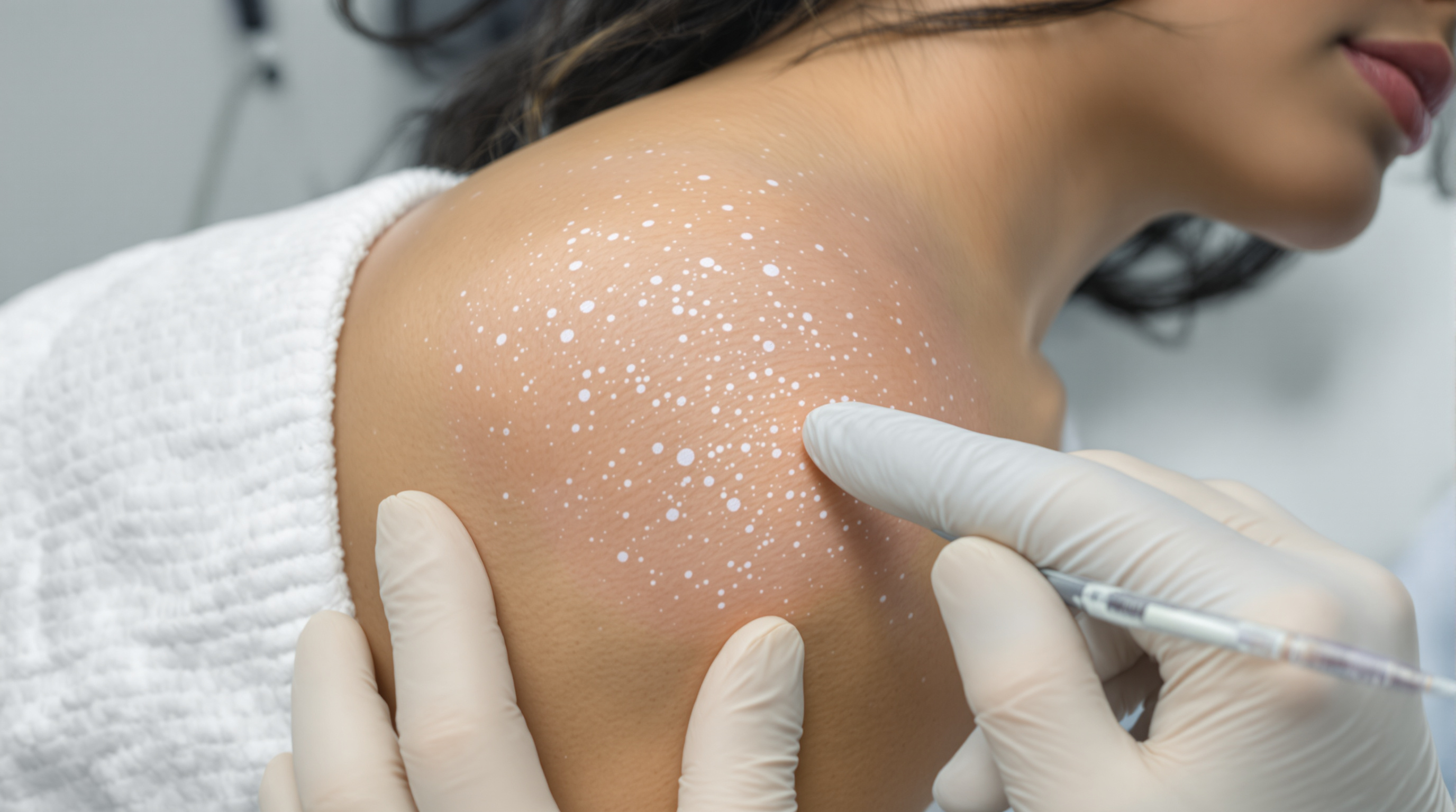
Patients who respond to vitiligo treatment often notice something odd before anything else: small brown dots appearing inside a white patch, each one centered on a hair. Over weeks these dots widen, merge, and eventually fill the patch. Dermatologists call this perifollicular repigmentation, and it is the most common pattern seen with phototherapy, topical calcineurin inhibitors, corticosteroids, and the newer JAK inhibitor creams. Understanding why it happens clarifies a great deal about which lesions are likely to recover and which are not.
The short version: in established vitiligo, the melanocytes of the surface epidermis in a patch are largely gone. The immune attack, driven mainly by CD8 positive T cells targeting melanocyte antigens, clears them out. What often survives is a separate population of melanocyte stem cells that sit deeper, in a protected niche of the hair follicle called the bulge region, near where the arrector pili muscle attaches. This zone is relatively shielded from immune surveillance, a property researchers describe as immune privilege. When treatment quiets the autoimmune attack, these stem cells can activate, divide, and send immature melanocytes migrating up the follicle and outward into the surrounding epidermis. The visible result is a pigmented dot at each follicular opening that slowly expands.
This mechanism has direct practical consequences.
Hair density predicts response. Skin rich in terminal hair follicles, such as the face, neck, and trunk, tends to repigment better because it holds more stem cell reservoirs per square centimeter. The lips, fingertips, knuckles, palms, soles, and genital mucosa have few or no follicles, which is a major reason acral and mucosal vitiligo are notoriously stubborn. It is not that the treatment fails to reach these sites. There is simply less raw material to rebuild from.
White hairs inside a patch are a warning sign. When the hairs within a vitiligo lesion have turned white, a condition called leukotrichia, it suggests the immune process has reached the follicular reservoir itself and destroyed or depleted the stem cells there. These patches respond poorly to medical therapy. This is one reason clinicians examine hair color inside lesions before setting expectations, and why longstanding, fully depigmented patches with white hair are often better candidates for surgical approaches such as melanocyte and keratinocyte cell suspension grafting, which physically transplants pigment cells from unaffected donor skin.
Repigmentation is slow by design. Stem cell activation, migration down and then across the epidermis, and maturation into pigment producing cells is a biological relay that takes months, not weeks. Most studies of narrowband UVB, tacrolimus, and topical ruxolitinib measure meaningful change at the 24 week mark and beyond, with continued improvement out to a year or more. A patient who sees no dots at six weeks has not necessarily failed treatment. Conversely, the appearance of even a few perifollicular dots is an encouraging early signal that the reservoir is intact and responding.
Not all repigmentation is follicular. Two other patterns exist. Marginal repigmentation creeps inward from the pigmented border of a patch, driven by melanocytes at the edge migrating laterally. Diffuse repigmentation, where the whole patch darkens evenly, is thought to involve surviving melanocytes scattered within the lesion or dermal reservoirs, and it is sometimes seen in newer lesions where destruction was incomplete. Mixed patterns are common. Some evidence suggests diffuse and marginal patterns can be less durable than follicular repigmentation, though data here are limited and individual results vary.
A related myth worth checking: the idea that light therapy "burns" pigment back into the skin. Phototherapy does not work by tanning the patch. Narrowband UVB acts through at least two mechanisms: it has local immunosuppressive effects that reduce the T cell attack, and it stimulates signaling molecules from keratinocytes, including endothelin 1 and various growth factors, that promote melanocyte stem cell activation and migration. This is why combining phototherapy with an anti inflammatory topical often outperforms either alone. One arm of the strategy stops the destruction, the other encourages repopulation.
Finally, the follicular reservoir concept explains why stability matters before surgical grafting. Transplanting melanocytes into skin where the autoimmune attack is still active is like reseeding a lawn during a drought. Most surgical protocols require the disease to have been stable, with no new or expanding lesions, for six to twelve months, sometimes supported by systemic therapy to hold the immune system in check.
The takeaway for anyone tracking their own treatment: look at the hairs. Pigmented hairs within a patch, especially on follicle dense skin, mean the reservoir is probably alive and the odds of medical repigmentation are meaningfully better. Tiny dots appearing around those hairs are the earliest visible proof the system is working. And patches on hairless or white haired skin deserve a frank conversation about combination therapy, longer timelines, or surgical options rather than more of the same. None of this replaces individualized evaluation by a qualified dermatologist, but it turns a confusing freckled pattern into what it actually is: the skin rebuilding itself from its deepest reserves.