
Explainer · July 16, 2026 · 5 min · By Verity Onwudiwe
Hypopigmented or Depigmented? How Clinicians Separate Pityriasis Alba From Early Vitiligo
Two of the most commonly confused pale patches in dermatology behave very differently under the skin. Here is how specialists tell them apart, and why the distinction changes everything about treatment.
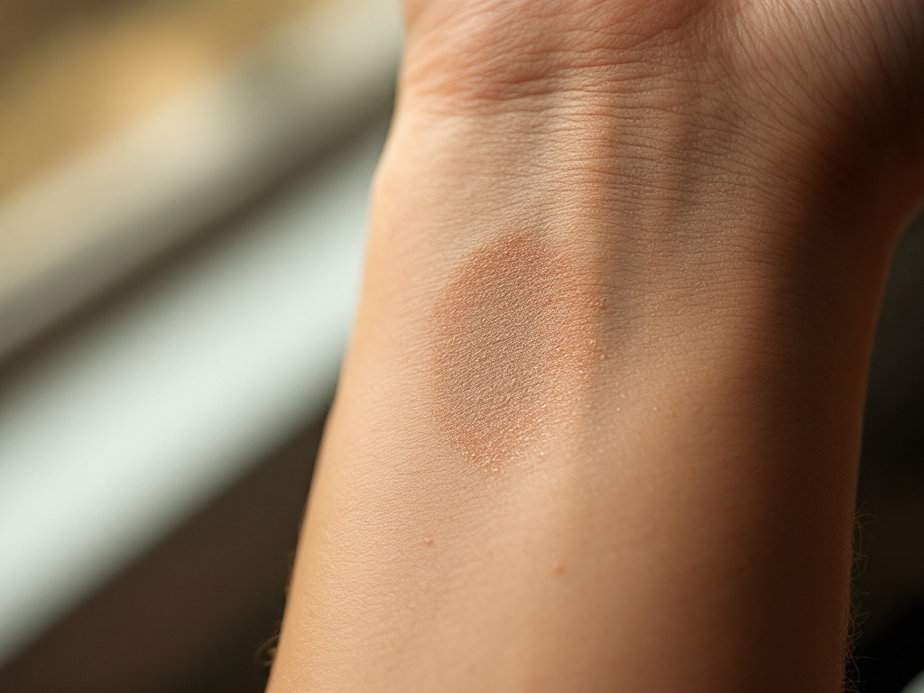
A pale patch on the face of a child, or on the arm of an adult after a summer tan, sends many families searching for answers. The two diagnoses that come up most often are pityriasis alba and vitiligo, and while they can look similar in a bathroom mirror, they are biologically distinct conditions with different causes, different trajectories, and very different treatment logic. Understanding the difference between a hypopigmented patch and a depigmented one is the single most useful concept for anyone navigating this territory.
The core distinction: less pigment versus no pigment. In pityriasis alba, melanocytes, the cells that produce pigment, are still present in the affected skin. They are simply producing less melanin and transferring it less efficiently to surrounding skin cells, usually because of low-grade inflammation associated with eczema-prone skin. The patch is hypopigmented: lighter than the surrounding skin, but not white. In vitiligo, the melanocytes themselves are progressively lost, most likely through an autoimmune process in which the body's own T cells target melanocyte proteins. The result is depigmentation: a patch that is truly white because there is no pigment left to see.
What each condition looks like in practice. Pityriasis alba typically appears as multiple faint, slightly scaly, poorly defined patches, most often on the cheeks, upper arms, and trunk of children and adolescents. The edges blur gradually into normal skin. The patches often become more noticeable in summer, not because they change, but because the surrounding skin tans and the contrast increases. Vitiligo patches tend to be sharply demarcated, milk-white, and smooth, without scale. They favor certain locations: around the eyes and mouth, the hands and fingertips, elbows, knees, and skin folds. Vitiligo can also show the Koebner phenomenon, in which new patches appear at sites of friction or injury, something pityriasis alba does not do.
The Wood's lamp test. The most reliable bedside tool for separating the two is a Wood's lamp, a handheld ultraviolet light used in a darkened room. Depigmented vitiligo skin fluoresces a bright, chalky blue-white because there is no melanin to absorb the UV light. Hypopigmented conditions like pityriasis alba show only a subtle accentuation, or none at all. This examination takes under a minute, is painless, and often settles the question without a biopsy. In people with very fair skin, where patches are hard to see in normal light, the Wood's lamp is frequently the only way to map the true extent of vitiligo.
Why the distinction matters for treatment. Pityriasis alba is fundamentally a mild eczema variant. It responds to moisturization, gentle skin care, and short courses of low-potency topical anti-inflammatories, and it typically resolves on its own over months to a few years as the skin barrier matures. Aggressive treatment is rarely warranted. Vitiligo, by contrast, requires suppressing an ongoing autoimmune attack and then coaxing surviving melanocytes, often those sheltering in hair follicles, to repopulate the skin. That is why vitiligo treatment leans on stronger tools: topical calcineurin inhibitors, topical JAK inhibitors, targeted narrowband UVB phototherapy, and in stable cases, surgical melanocyte transfer. Treating vitiligo with the gentle approach appropriate for pityriasis alba wastes the early window when repigmentation is easiest, and treating pityriasis alba with vitiligo-strength therapy exposes a child to unnecessary risk.
Other look-alikes worth knowing. The differential does not end with these two. Tinea versicolor, a common yeast overgrowth, causes hypopigmented patches with fine scale, usually on the chest and back, and shows a yellow-green tint under Wood's lamp. Post-inflammatory hypopigmentation follows a rash, burn, or procedure and fades as melanocytes recover. Nevus depigmentosus is a stable, congenital pale patch present from infancy that grows proportionally with the child and never spreads the way vitiligo can. A clinician sorting through these options relies on history, distribution, texture, and the Wood's lamp far more than on any single glance.
Red flags that point toward vitiligo. Certain features should prompt a prompt dermatology evaluation rather than watchful waiting: patches that are stark white rather than faintly pale, sharp borders, involvement of the hands, lips, or genitals, white hairs growing within a patch, new spots appearing at sites of scrapes or friction, or a personal or family history of thyroid disease or other autoimmune conditions. Early vitiligo, particularly on the face, responds far better to treatment than long-established disease, so timing genuinely matters.
The takeaway is straightforward. Pale is not the same as white, and less pigment is not the same as no pigment. A careful examination, often with nothing more high-tech than an ultraviolet lamp in a dark room, can distinguish a benign, self-resolving condition from an autoimmune one that rewards early intervention. Anyone with a persistent or spreading pale patch deserves that examination rather than a guess.