
Explainer · July 30, 2026 · 4 min · By Verity Onwudiwe
Is That White Patch Vitiligo? How Clinicians Separate It From Four Common Look-Alikes
Not every pale patch of skin is vitiligo. Here is how dermatologists actually distinguish true depigmentation from pityriasis alba, tinea versicolor, post-inflammatory hypopigmentation, and idiopathic guttate hypomelanosis.
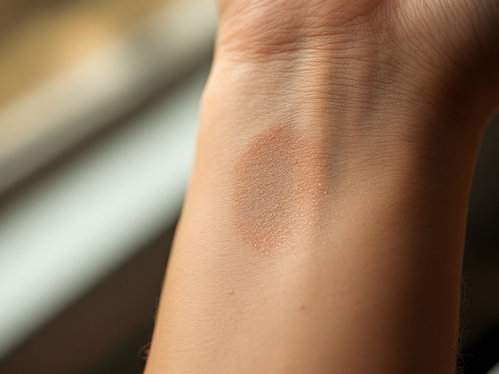
A new white or light patch on the skin sends many people straight to a search engine, and the first result is often vitiligo. But dermatologists see several conditions every week that mimic it, and the distinction matters. Vitiligo is an autoimmune process that destroys melanocytes, the pigment-producing cells, while most look-alikes leave those cells alive but underperforming. Different mechanism, different prognosis, different treatment.
The core distinction: depigmentation versus hypopigmentation. In vitiligo, melanocytes in the affected patch are largely gone, so the skin loses essentially all pigment. The patch tends to be milk white or chalk white with fairly crisp borders. In hypopigmentation, melanocytes remain but produce less melanin, so patches look off-white, pale tan, or simply lighter than surrounding skin, often with softer, blurrier edges. This is why the first tool a clinician reaches for is usually a Wood's lamp, a handheld ultraviolet light used in a darkened room. Fully depigmented vitiligo skin fluoresces a bright blue-white because there is no melanin left to absorb the light. Hypopigmented patches show only a modest accentuation. It is a simple, painless test, and it changes the diagnosis more often than patients expect.
Look-alike one: pityriasis alba. This is extremely common in children and teens, especially on the cheeks and upper arms, and especially in people with a history of eczema or dry skin. The patches are pale rather than white, often slightly scaly or rough, and the borders fade gradually into normal skin. Mechanistically, low-grade eczematous inflammation temporarily disrupts the transfer of melanin from melanocytes to surrounding skin cells. The melanocytes survive. Pityriasis alba typically improves with moisturizers, gentle skin care, sometimes a short course of a mild topical anti-inflammatory, and time. It does not progress to vitiligo.
Look-alike two: tinea versicolor. This is caused by an overgrowth of Malassezia, a yeast that lives on everyone's skin. On darker or tanned skin it produces lighter patches, usually on the chest, back, and shoulders, often with a fine, dust-like scale that becomes visible when the skin is stretched or scraped. The yeast produces azelaic acid and related compounds that inhibit tyrosinase, the key enzyme in melanin production, which is why the patches lighten. Under a Wood's lamp it may show a yellowish or coppery glow rather than the stark blue-white of vitiligo. Antifungal washes or creams clear the yeast, though the pigment can take weeks to months to catch up, which sometimes leads people to believe the treatment failed.
Look-alike three: post-inflammatory hypopigmentation. Skin that has been inflamed or injured, whether by eczema, psoriasis, a burn, a laser treatment, or even an aggressive chemical peel, can heal lighter than the surrounding skin. Again, the melanocytes are usually stressed rather than destroyed, and pigment often returns over months as the skin recovers. The history is the giveaway: the pale area maps precisely onto a site of prior rash or trauma. True vitiligo can also appear at sites of skin injury through a phenomenon called the Koebner response, which is one reason a careful history and follow-up matter. A patch that keeps expanding beyond the original injury, or turns fully white under Wood's lamp examination, deserves reassessment.
Look-alike four: idiopathic guttate hypomelanosis. These are small, flat, well-defined white spots, usually 2 to 6 millimeters across, scattered on the shins and forearms of adults, and they become more common with age and cumulative sun exposure. They are stable, harmless, and do not merge into larger patches the way active vitiligo can. The mechanism appears to involve a localized age-related and photodamage-related decline in melanocyte number and function. No treatment is medically necessary, and sun protection helps limit new spots.
Why getting this right changes what happens next. Vitiligo is autoimmune, so its management aims at calming the immune attack and coaxing surviving melanocytes, often those hiding in hair follicles, to repopulate the skin. Options include topical corticosteroids, topical calcineurin inhibitors, narrowband UVB phototherapy, and more recently a topical JAK inhibitor approved for nonsegmental vitiligo, which blocks the interferon-gamma signaling pathway that drives melanocyte destruction. None of those approaches is appropriate for a yeast overgrowth or a resolving eczema patch, and using them there wastes time and money.
What patients can watch for. Features that make vitiligo more likely include chalk-white color, sharply defined borders, symmetry on both sides of the body, involvement of the face, hands, or around body openings, whitening of hairs within the patch, and steady enlargement over weeks to months. Features favoring a mimic include scale, itch, a history of rash or injury in that exact spot, and color that is pale rather than truly white.
The practical takeaway: a pale patch is a reason for a proper skin exam, not panic. A Wood's lamp, a good history, and occasionally a scraping or biopsy resolve most of these cases in a single visit, and the majority of white patches walking into clinics are not vitiligo at all.
Related reading: White hairs inside a vitiligo patch: the sign that quietly predicts how well it will repigment.